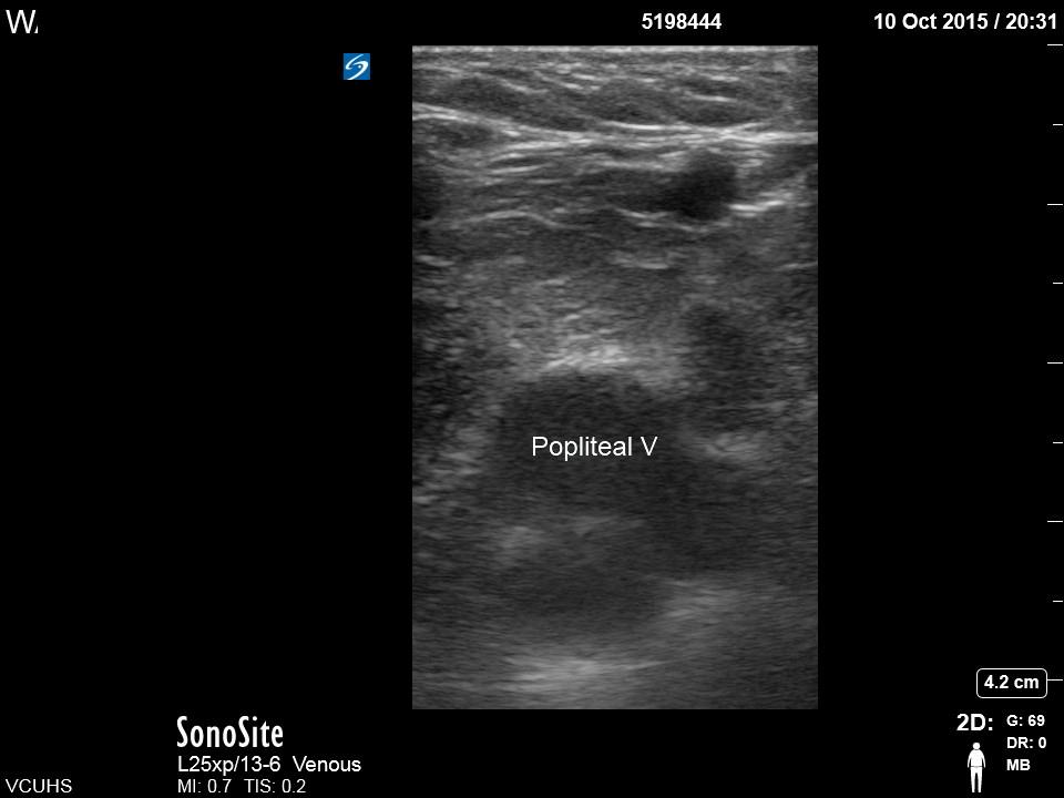
Pearls for obtaining a good DVT study, as demonstrated by Dr. Wilson’s case
- Here is the image that Dr. Wilson caught on his exam. Notice the inability to compress the vein sealing the diagnosis.
Complete study:
Limited Bedside Venous Ultrasound

A. Clip of popliteal trifurcation with compression
B. Clip of popliteal vein with compression

C. Clip of Saphenous Vein joining Common Femoral Vein with compression

D. Clip of Femoral Vein joining Deep Femoral Vein with compression

E. If it is visible, continue to follow the Femoral Vein distally doing serial compression.
